Materials characterization Unit
Scientific equipment:
X-ray diffractometer SmartLab (Rigaku, Japan, 2011): 9 kW x-ray tube with rotating Cu anode; optics: parallel beam/Bragg-Brentano, polycapillary; Ge monochromators: Ge (220)´2, Ge (220)´4, Ge (400)´2; analysers: Ge (220)´2, Ge (400)´2; bent graphite monochromator for diffracted beam; CALSA – crystal array monochromator of especially high resolution and intensity for diffracted beam; detectors: scintillation SC-70, 1D (linear) D/teX Ultra, 2D (two dimensional) Pilatus 100K; sample stages: Eulerian cradle (Y, f, x, y, z), X-Y (50-50 mm) stage, RxRy stage, stage for powder samples, high temperature stage Anton Paar DHS 1100 (temperatures up to +1100 °C in vacuum or inert gas atmosphere); vacuumed path for x-rays diffracted at small angles (SAXS); special axis for In-plane method; powder diffraction database PDF4+ (2011 release).
X-ray diffractometer D8 Advance (Bruker AXS, Germany, 2003 m.): 2,4 kW sealed x-ray tube with Cu anode; optics: parallel beam/Bragg-Brentano; V grove Ge monochromator; scintillation detector; Eulerian cradle (Y, f, x, y, z); powder diffraction database PDF2 (2003 release).
Fluorescent x-ray spectrometer with wave dispersion (WDXRF) Axios mAX (Panalytical, Netherlands, 2011). 4 kW x-ray tube with Rh anode STmax160; detectors: proportional flow, proportional sealed (with Xe gas), highly effective scintillation; six crystals-analysers (elements from O to U); software: SuperQ – for standard-less quantitative analysis; Ni-Fe-Co – for quantitative analysis of alloys; FP-Multi – for quantitative analysis and determination of thickness of thin films; Pro-Trace – for quantitative analysis of trace elements.
Scanning electron microscope Helios NanoLab 650 (FEI, Netherlands, 2011) with Schottky type field emission electron source, galium ion source, TEM sample lift-out mechanism Omniprobe 100.7 (Oxford Instruments), platinum deposition and selective carbon mill gas injection systems, cryocleaner and plasma cleaner. X-ray spectrometer INCAEnergy (Oxford Instruments) with X-Max detector. ThinFilm ID software for thin film composition and thickness determination. High tension to 30 kV, resolution: 0,8 nm (30-2 kV); 0,9 nm (1 kV); 1,5 nm (200 V).
Scanning electron microscope EVO-50 EP (Carl Zeiss SMT, Germany, 2006) with tungsten and LaB6 electron sources, secondary, VP secondary and backscattered electron detectors, energy and wave dispersion (EDS and WDS) X-Ray spectrometers INCA (Oxford Instruments), high (about 10-3 Pa) and low (5- 2000 Pa) vacuum in a sample chamber. High tension to 30 kV, resolution 2 nm (LaB6), 3 nm (W) at 15 kV.
Transmission electron microscope Tecnai G2 F20 X-TWIN (FEI, Netherlands, 2011) with Schottky type field emission electron source. Flexible high tension: 20, 40, 80, 120, 160, 200 kV. High angle annular dark field (HAADF) detector, energy dispersive x-ray spectrometer EDAX with r-TEM detector, 11 MPix ORIUS SC1000B (Gatan) CCD camera, single and double tilt specimen holders, sample plasma cleaner. Resolution (point, line) – (0,25-0,102 nm).
Spectrometer of x-ray photoelectrons and Auger electrons ESCALAB MK II (VG Scientific, United Kingdom, 1985).
Stationary optic microscope Neophot 2 (Carl Zeiss Jena, Germany, 1985).
Setup for spatially-resolved photoluminescence study. The equipment consists of multi-modular microscopy system WITec Alpha 300 capable for operation in i) confocal, ii) scanning near-field optical (SNOM), and iii) atomic force microscopy modes. Photoluminescence is recorded using spectrometer UHTS 300 coupled with CCD camera or photomultiplier Hamamatsu H8259-01. High spatial resolution is ensured by NIKON objective of large numerical aperture (NA=0.9).
Equipment for sample preparation:
Pulverisette 6 (FRITSCH, Germany) – the ball grinding-mill for a preparation of powder samples (salts, soil, ceramics, minerals) for WD-XRF and XRD measurements. Volume of tungsten carbide bowl 80 ml, 5 balls of tungsten carbide;
TP-20 (Herzog, Germany) Hydraulic press for the formation powder samples in the form of tablet (F 37 mm);
Eagon 2 (Panalytical, Netherlands, 2011) – fully automated fusion furnace for fused bead sample preparation (F 27 mm);
Tegramin-25 (Stuers, Denmark) – fully automated system for sample preparation by surface polishing.
Techniques:
XRD investigations of polycrystalline materials:
- Qualitative and quantitative phase analysis;
- Qualitative and quantitative phase analysis of micro-quantities using polycapillary optics;
- XRD examination at a high temperature (from +20 to +1100 °C in vacuum or inert gas atmosphere);
- Determination of crystallite size, micro and macro stresses, preferred crystallographic orientation using pole figures;
- Investigations of thin films by in-plane technique;
- Investigations of epitaxial layers by rocking curves and reciprocal lattice mapping techniques;
- Determination of thickness, density and roughness of thin layer and multi-layers by x-ray reflectometry (XRR);
- Determination of size distribution of nanoparticles and nanopores by small angle scattering of x-ray (SAXS) in transmission and reflection modes.
Qualitative and quantitative chemical analysis of powders, minerals, soil, catalysts, ceramics, glass, plastics, metals and alloys using WDXRF, electron microprobe analysis, x-ray photoelectrons and Auger elektronas spectroscopy
Investigation of surface morphology using scanning electron microscopy (SEM amd FE-SEM).
Investigation of cross section using focussed ion beam (FIB) and field emission scanning electron microscopy (FE-SEM).
Investigation of internal structure by transmission electron microscopy (TEM, EDX, EDS).
Atomic force microscopy.
Surface and metallographic investigation by optical microscopy.
Pholuminescence spectroscopy with subwavelength spatial resolution
Product testing for fulfilment of RoHS requirements, qualitative and quantitative determination of hazardous materials.
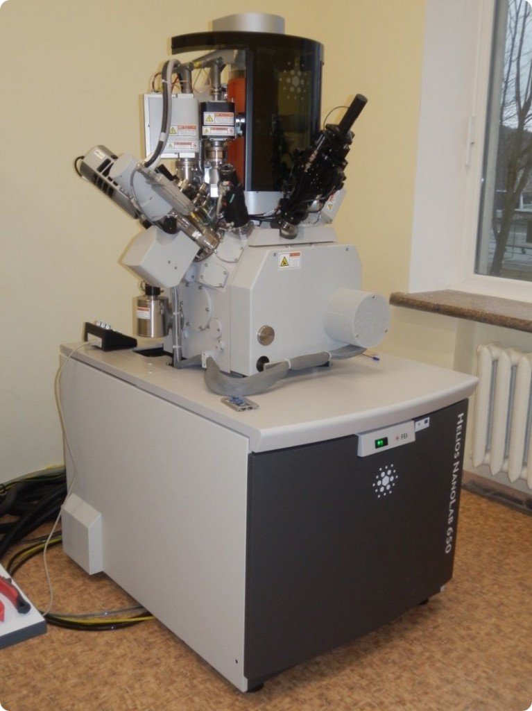
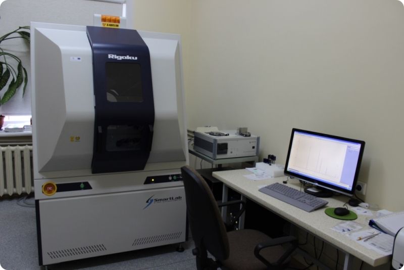
Dual beam system (FE-SEM + FIB) Helios Nanolab 650 X-ray diffractomer SmartLab (Rigaku) with 9 kW rotating anode.
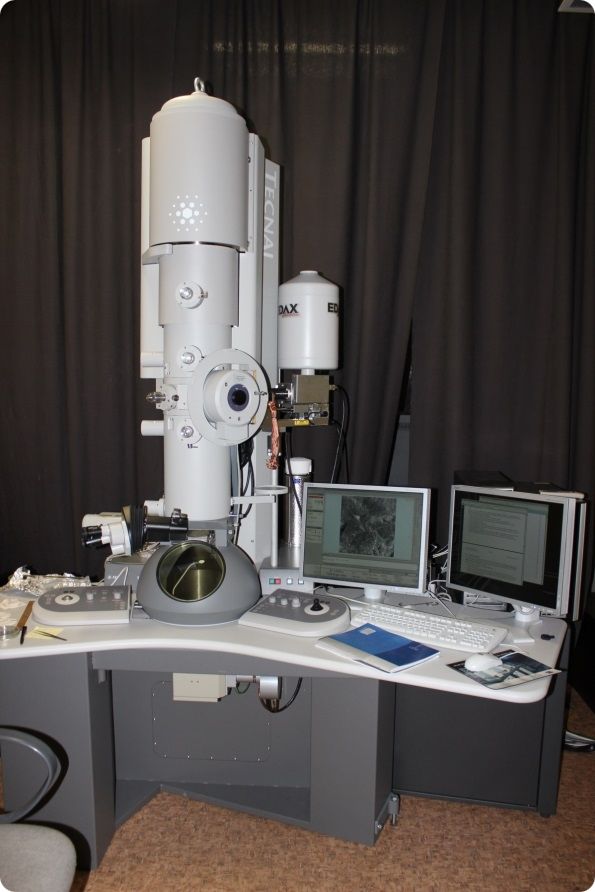
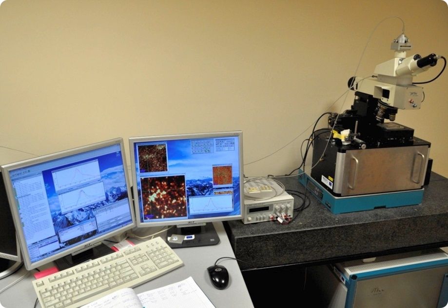
Transmission electron microscope Tecnai G2 F20 X-TWIN WITec Alpha 300 operatiing in confocal, scanning near-field optical (SNOM), and atomic force microscopy modes